
Calcium sulphate as a local antibiotic carrier in the treatment of bone infections and deep wounds
When a resorbable antibiotic carrier has a role alongside surgical debridement, what the evidence shows and how to care for the wound after surgery.
Oliver Jurić · 10 min read
English translation of the original Croatian article; the Croatian version is authoritative. Educational content only — not individual diagnosis or treatment.
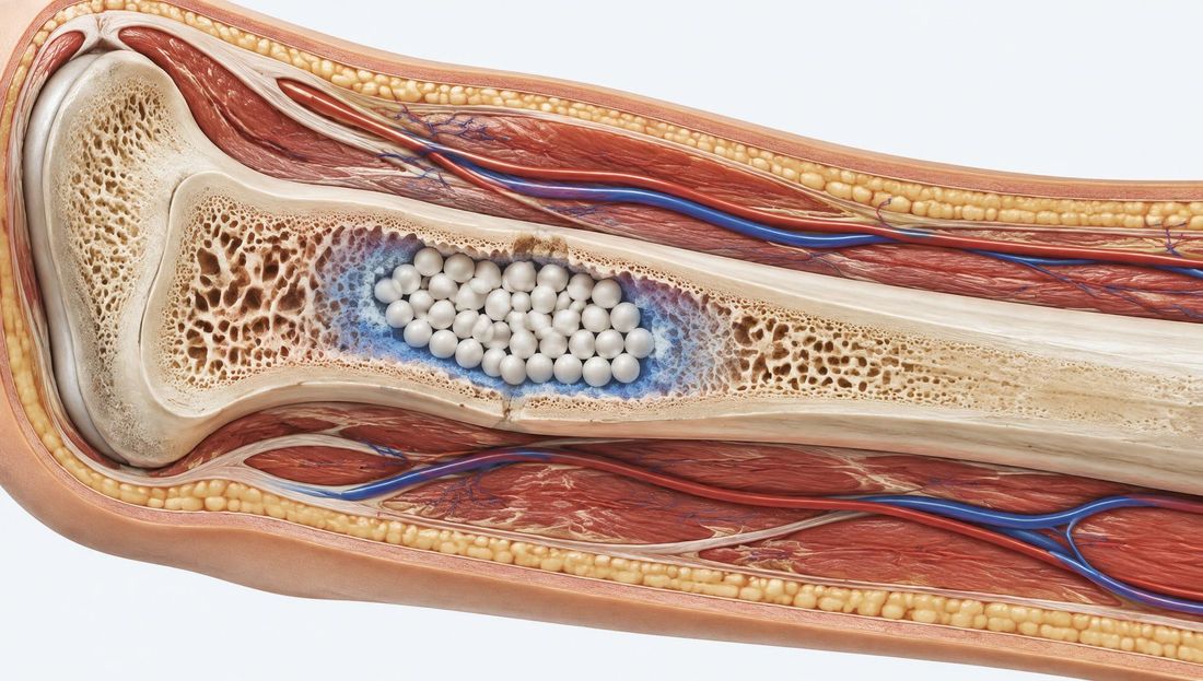
Resorbable calcium sulphate releases antibiotics directly into the bone defect after debridement. It complements surgical and systemic treatment, and the most common adverse effect is prolonged serous wound discharge.
Ključne točke
- Synthetic calcium sulphate is a resorbable carrier that releases antibiotics directly into a bone defect or deep cavity after surgical debridement.
- A local carrier complements, rather than replaces, radical debridement, stabilisation, soft-tissue coverage and targeted systemic antibiotic therapy.
- Unlike polymethylmethacrylate (PMMA) cement beads, calcium sulphate is resorbed and generally does not require a second procedure for removal.
- The most common adverse effect is prolonged serous wound discharge, which does not necessarily indicate infection but requires assessment and effective protection of the periwound skin.
- The evidence is largely observational; results in diabetic foot osteomyelitis are inconsistent, and use requires caution.
Why use local antibiotic therapy?
Chronic osteomyelitis, fracture-related infection and infection around orthopaedic implants share a common feature: microorganisms live in poorly perfused or necrotic tissue and in biofilms. Systemically administered antibiotics reach these areas at low concentrations, whereas activity against bacteria in biofilms requires concentrations many times higher than the usual minimum inhibitory concentration (MIC).
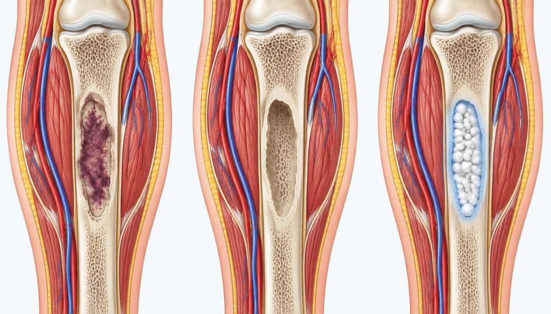
A local antibiotic carrier is placed in the cavity created by surgical removal of necrotic bone and infected tissue. The aim is to achieve a high local drug concentration precisely where the risk of recolonisation is greatest, with low systemic exposure. The carrier also fills dead space, which would otherwise fill with haematoma and provide a favourable environment for recurrent infection.

What is calcium sulphate as a carrier?
High-purity synthetic calcium sulphate, rather than natural gypsum, is used for this indication. The powder is mixed with liquid and the selected antibiotic, then formed into beads or a paste, or injected into the defect. After setting, the carrier gradually dissolves in the tissue over several weeks, releasing the antibiotic as it does so.
Properties relevant to clinical practice
- Resorption: the carrier breaks down in the tissue, so a second procedure for its removal is generally unnecessary.
- Release profile: most of the antibiotic is released in the first few days, with lower release over the following weeks; the exact kinetics depend on the drug, the form of the carrier and local conditions.
- Osteoconductivity: the carrier can act as a temporary scaffold for bone ingrowth, but complete filling of the defect with bone tissue is not the rule.
- Antibiotic compatibility: heat generation during setting is considerably lower than with bone cement, allowing heat-sensitive drugs to be used.
In laboratory models, antibiotic-loaded calcium sulphate beads prevented colonisation and biofilm formation on implant surfaces and were also active against established biofilms. These findings explain the mechanism but do not demonstrate clinical outcomes.
Comparison with non-resorbable carriers
The conventional local carrier consists of antibiotic-loaded polymethylmethacrylate beads or spacers. These are well established and mechanically stable, but they do not degrade and are usually removed in a second procedure. Once the antibiotic has been released, their surfaces can provide a substrate for biofilm formation.
| Characteristic | Calcium sulphate | Polymethylmethacrylate (PMMA) |
|---|---|---|
| Degradation in tissue | Yes, over several weeks | No |
| Need for removal | Generally no | Usually yes, in a second procedure |
| Heat generation during setting | Low | High (exothermic reaction) |
| Antibiotic choice | Broader, including heat-sensitive drugs | Limited to heat-stable drugs |
| Mechanical load-bearing capacity | None | Limited; a spacer can temporarily maintain space |
| Most common local problem | Serous wound discharge | Residual foreign material and the need for reoperation |
Tablicu je na užim zaslonima moguće pomicati vodoravno.
In a small randomised study of chronic osteomyelitis and infected nonunion, both carriers achieved similar infection eradication rates, while the resorbable carrier group underwent fewer repeat operations. The study included 30 patients and should therefore be interpreted as preliminary evidence.
When is use considered?
- Chronic osteomyelitis after surgical removal of necrotic bone.
- Fracture-related infection and infected nonunion, as part of staged treatment.
- Periprosthetic joint infection, during revision surgery and as an adjunct to other measures.
- Selected cases of diabetic foot osteomyelitis after surgery, following a multidisciplinary decision.
- Deep soft-tissue cavities after debridement of infected tissue, when dead space needs to be filled.
Debridement is a prerequisite
A local carrier does not treat infection that the surgeon has not removed. The outcome depends primarily on complete removal of necrotic bone, sequestra and infected foreign bodies, bone stability and the ability to cover the defect with healthy, well-perfused soft tissue. In chronic limb ischaemia, the feasibility of revascularisation should be assessed first, because without a blood supply neither a local nor a systemic approach can achieve healing.
Bone and deep-tissue samples for microbiological and histopathological analysis are taken before the carrier is placed. The results guide systemic antibiotic therapy and allow assessment of whether the chosen local antibiotic was appropriate.
Antibiotic selection
Vancomycin, gentamicin and tobramycin are the most commonly used antibiotics, alone or in combination, because together they cover a large proportion of Gram-positive and Gram-negative pathogens. The drug must be suitable for mixing with the carrier and must not adversely alter its setting time. Some antibiotics delay or prevent setting, so the combination and dose are selected according to the carrier's instructions and verified compatibility data.
Selection should take account of the known or expected pathogen, previous microbiological findings, the patient's drug hypersensitivity and renal function. Local administration does not remove the need for culture-directed systemic therapy.
Monitoring and adverse effects
Wound discharge
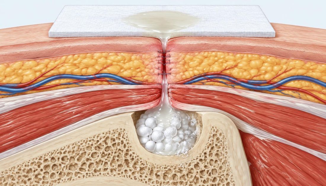
As the carrier dissolves in the tissue, fluid forms and may drain through the surgical wound, particularly when a large volume of carrier has been used or the soft-tissue layer is thin. In a large series of chronic osteomyelitis cases, prolonged discharge lasting more than two weeks was recorded in 15.4% of cases and was not associated with recurrent infection. In a systematic review and meta-analysis, wound complications were recorded in 17% of patients treated for osteomyelitis.
The discharge may be clear or whitish and milky because of the dissolved carrier. It should be distinguished from purulent discharge, spreading erythema, increasing pain and fever, which indicate infection. For wound care, this means more frequent assessment, an absorbent dressing appropriate to the volume of exudate and protection of the periwound skin from maceration.
Systemic effects
- Transient hypercalcaemia has been reported, particularly with larger carrier volumes; serum calcium levels are monitored in patients at risk.
- Renal function is monitored when aminoglycosides or vancomycin are used, particularly in patients with pre-existing chronic kidney disease, older people and those receiving other nephrotoxic drugs.
- Heterotopic ossification and soft-tissue irritation have rarely been reported, mainly when the carrier has not remained confined to the bone defect.
Razina dokazaLow to moderatePedrazzini et al., systematic review and meta-analysis, 2026.Included 41 studies, with no randomised controlled trials.
What does the evidence show, and what are its limitations?
A 2026 systematic review and meta-analysis included 41 studies and found no randomised controlled trials. The pooled infection eradication rate was 88% for osteomyelitis and also 88% for periprosthetic joint infection, with substantial heterogeneity between studies. These results reflect the overall treatment rather than the effect of the carrier alone, as patients also underwent surgery and received systemic antibiotics.
Data on diabetic foot osteomyelitis are sparse and inconsistent. In a retrospective cohort of 45 patients, 29% were ulcer-free at six months, osteomyelitis recurred in 42%, wound discharge was recorded in 62% and acute kidney injury in 9%. The authors recommended caution pending randomised studies.
In their joint 2023 guidelines, the International Working Group on the Diabetic Foot (IWGDF) and the Infectious Diseases Society of America (IDSA) suggest not using local antibiotics as an adjunct to systemic therapy for soft-tissue and bone infections of the diabetic foot. This is because the certainty of the evidence is low: available studies have a high risk of bias and inconsistent results, and the safety of routine use has not been clearly established. The use of a local carrier in this population is therefore an individual decision for an experienced team, rather than standard practice.
Role in wound care
For the wound-care team, most of the work begins after surgery. It is important to know that a resorbable carrier has been placed in the wound, what discharge to expect and which signs indicate infection or compromised wound closure.
- Document the type of carrier, the added antibiotic, the date of placement and the anatomical site.
- At each dressing change, describe the amount, colour and odour of the discharge and the condition of the wound edges and periwound skin.
- Select a dressing according to the volume of exudate and protect the periwound skin; for heavy discharge, consider more frequent dressing changes or a more absorbent dressing system.
- Do not irrigate the wound under pressure or remove white material appearing in the wound without consulting the operating surgeon.
- Monitor laboratory results requested by the operating surgeon, such as renal function and calcium levels.
- Notify the operating surgeon without delay if there is purulent discharge, spreading erythema, increasing pain, fever or separation of the wound edges.
Frequently asked questions
Česta pitanja
Does white or milky discharge after surgery indicate infection?
Not necessarily. The dissolved carrier can produce clear or milky discharge that may persist for several weeks. Purulent discharge, malodour, spreading erythema, increasing pain and fever indicate infection.
Does the carrier need to be removed in a second procedure?
Generally not, because calcium sulphate is resorbed in the tissue. A repeat procedure may be necessary because of the course of the infection itself or wound problems, rather than to remove the carrier.
Can a local carrier replace systemic antibiotics?
No. A local carrier complements surgical treatment and culture-directed systemic therapy.
Is use justified in diabetic foot osteomyelitis?
The evidence is limited and inconsistent, and the international IWGDF/IDSA guidelines suggest not using local antibiotics routinely. The decision is individualised and made by a multidisciplinary team.
Izvori
References
Popis izvora korištenih pri pisanju ovog članka.
1. izvor: McKee MD, Wild LM, Schemitsch EH, Waddell JP. The use of an antibiotic-impregnated, osteoconductive, bioabsorbable bone substitute in the treatment of infected long bone defects: early results of a prospective trial. J Orthop Trauma. 2002;16(9):622–627.
DOI: 10.1097/00005131-200210000-00002
Otvori izvor (vanjska poveznica)2. izvor: McKee MD, Li-Bland EA, Wild LM, Schemitsch EH. A prospective, randomized clinical trial comparing an antibiotic-impregnated bioabsorbable bone substitute with standard antibiotic-impregnated cement beads in the treatment of chronic osteomyelitis and infected nonunion. J Orthop Trauma. 2010;24(8):483–490.
DOI: 10.1097/BOT.0b013e3181df91d9
Otvori izvor (vanjska poveznica)3. izvor: Beuerlein MJ, McKee MD. Calcium sulfates: what is the evidence? J Orthop Trauma. 2010;24(Suppl 1):S46–S51.
DOI: 10.1097/BOT.0b013e3181cec48e
Otvori izvor (vanjska poveznica)4. izvor: Ferguson JY, Dudareva M, Riley ND, Stubbs D, Atkins BL, McNally MA. The use of a biodegradable antibiotic-loaded calcium sulphate carrier containing tobramycin for the treatment of chronic osteomyelitis: a series of 195 cases. Bone Joint J. 2014;96-B(6):829–836.
DOI: 10.1302/0301-620X.96B6.32756
Otvori izvor (vanjska poveznica)5. izvor: Howlin RP, Brayford MJ, Webb JS, et al. Antibiotic-loaded synthetic calcium sulfate beads for prevention of bacterial colonization and biofilm formation in periprosthetic infections. Antimicrob Agents Chemother. 2015;59(1):111–120.
DOI: 10.1128/AAC.03676-14
Otvori izvor (vanjska poveznica)6. izvor: McConoughey SJ, Howlin RP, Wiseman J, Stoodley P, Calhoun JH. Comparing PMMA and calcium sulfate as carriers for the local delivery of antibiotics to infected surgical sites. J Biomed Mater Res B Appl Biomater. 2015;103(4):870–877.
DOI: 10.1002/jbm.b.33247
Otvori izvor (vanjska poveznica)7. izvor: Laycock PA, Cooper JJ, Howlin RP, et al. In Vitro Efficacy of Antibiotics Released from Calcium Sulfate Bone Void Filler Beads. Materials (Basel). 2018;11(11):2265.
DOI: 10.3390/ma11112265
Otvori izvor (vanjska poveznica)8. izvor: Alkayali T, Casciato D, Wynes J, Chua J, Doub JB. Are Biodegradable Calcium Sulfate Antibiotic Beads Effective and Safe Adjuvants for Diabetic Foot Osteomyelitis? Cureus. 2024;16(1):e52444.
DOI: 10.7759/cureus.52444
Otvori izvor (vanjska poveznica)9. izvor: Senneville É, Albalawi Z, van Asten SA, et al. IWGDF/IDSA guidelines on the diagnosis and treatment of diabetes-related foot infections (IWGDF/IDSA 2023). Diabetes Metab Res Rev. 2024;40(3):e3687.
DOI: 10.1002/dmrr.3687
Otvori izvor (vanjska poveznica)10. izvor: Pedrazzini A, Suravaram V, Lu VYZ, Hoch A, Zingg PO, Andronic O. Outcomes and safety of antibiotic-loaded calcium sulfate beads in orthopaedics: A systematic review and meta-analysis. J Orthop. 2026;76:359–376.
DOI: 10.1016/j.jor.2026.04.019
Otvori izvor (vanjska poveznica)