
Chronic wound infection: diagnosis and antimicrobial therapy
How to distinguish colonisation from infection, understand biofilm and use antiseptics and antibiotics rationally.
Oliver Jurić · 23 min read
English translation of the original Croatian article; the Croatian version is authoritative. Educational content only — not individual diagnosis or treatment.
Chronic wound infection is a clinical diagnosis, not a microbiological finding. Safe treatment requires differentiation between colonisation and local, spreading and systemic infection, assessment of perfusion and the depth of the process, appropriate source control and antimicrobial therapy only when clearly indicated. This approach protects patients both from undertreatment of serious infection and from unnecessary use of antimicrobial agents.
Ključne točke
- All open chronic wounds contain microorganisms; their presence alone does not establish infection.
- Infection is primarily diagnosed clinically, based on the host response and signs of tissue invasion, while microbiology helps identify the causative organism and guide treatment.
- The term ‘critical colonisation’ is no longer recommended; the continuum distinguishes contamination, colonisation, local infection, spreading infection and systemic infection.
- Biofilm is important, but it must not be equated with clinical infection, nor should a reduction in bioburden be presented as evidence that infection has been cured.
- A clinically uninfected wound does not require a systemic antibiotic solely to accelerate healing or prevent infection.
- Antimicrobial stewardship means using antimicrobial therapy only when needed, in the most appropriate form, with a sufficiently narrow spectrum and only for as long as clinically justified.
Wound infection begins with the relationship between microorganisms and the host
The most important conceptual error in chronic wound care is equating bacteria with infection. An open wound very quickly comes into contact with microorganisms and may remain colonised throughout the healing process. Infection develops when microorganisms proliferate and invade tissue to an extent that triggers a host response and tissue damage. The diagnosis of infection therefore cannot be based solely on a positive swab, a bacterial count or the presence of a species considered a potential pathogen. The European Wound Management Association (EWMA) document published in 2022 cautions that the heterogeneous distribution of microorganisms within a wound prevents infection from being reliably defined by a single quantitative threshold.
The International Wound Infection Institute (IWII) wound infection continuum, updated in 2022, distinguishes five conceptual stages: contamination, colonisation, local infection, spreading infection and systemic infection. The term ‘critical colonisation’ was removed because it is not possible to identify a universal point at which the microbial burden suddenly becomes ‘critical’. Instead, local infection in chronic wounds may initially produce subtle signs, with classic manifestations of inflammation appearing only later.
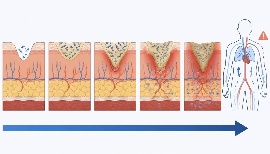

As the continuum diagram shows, the transition between stages is neither instantaneous nor clear-cut: the balance between microbial burden, virulence and host resistance changes, and clinical decisions follow that balance, not an isolated laboratory value.
| Clinical state | Relationship between microorganisms and the host | Typical clinical findings | Antimicrobial treatment |
|---|---|---|---|
| Contamination | Microorganisms present without significant proliferation | No clinical host response | Not indicated solely because microorganisms are present |
| Colonisation | Limited proliferation without a significant host response | No clinical infection or delayed healing attributable to infection | Routine antimicrobial treatment is not justified |
| Local infection | Proliferation triggers a local host response | Subtle or classic local signs | Local wound management and source control; antimicrobial strategy according to the clinical situation |
| Spreading infection | The process extends beyond the immediate wound area | Cellulitis, progressive induration or erythema, lymphangitis, deeper involvement | Systemic assessment and systemic antimicrobial therapy as indicated |
| Systemic infection | Infection triggers a systemic response | General deterioration, sepsis, organ dysfunction | Emergency hospital care and treatment according to infection severity |
Tablicu je na užim zaslonima moguće pomicati vodoravno.
IWII describes local infection as a process confined to the wound and its immediate surroundings. Subtle signs include hypergranulation, friable granulation tissue that bleeds, pocketing or epithelial bridging, increased exudate and an unexpected stall in healing. Classic signs include new or increased pain, warmth, swelling, purulent discharge, wound enlargement and increased malodour. No individual sign is sufficiently reliable to confirm or exclude infection on its own.
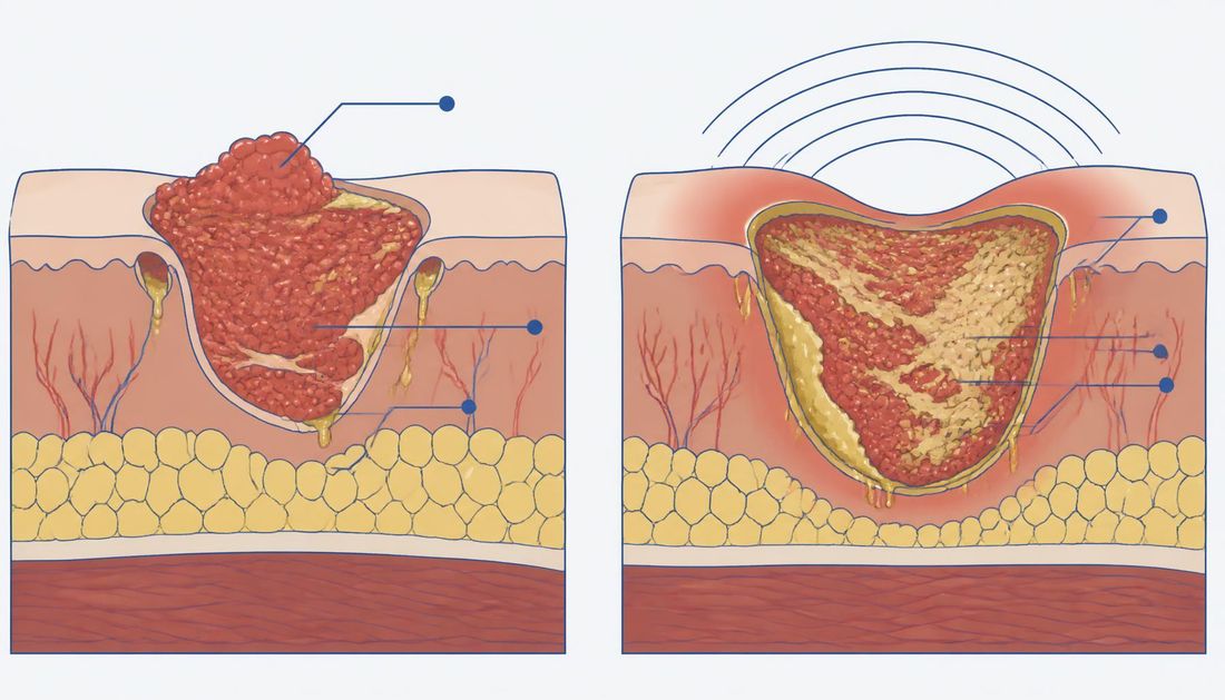
The comparison of the two cross-sections illustrates the key point for practice: the subtle presentation on the left belongs to the same diagnostic category as the classic presentation on the right, so decisions must not be delayed simply because the wound does not look dramatic.
Clinical assessment: infection may be overt or concealed
Classic inflammatory signs may be less pronounced in older people, immunocompromised patients and people with diabetes, neuropathy or severely impaired perfusion. A quiet-looking wound is therefore not sufficient to exclude infection when accompanied by unexpected deterioration, new pain, increased exudate, friable granulation tissue or stalled healing. IWII emphasises the need to combine several signs and symptoms rather than rely on a single finding.
The assessment must also address another question: is there a condition that could mimic infection? Venous dermatitis can cause redness and exudation. Acute lipodermatosclerosis can be painful and erythematous. Contact dermatitis may develop after the use of topical products. Inflammatory conditions such as pyoderma gangrenosum may worsen after aggressive debridement. Ischaemia can cause pain, necrosis and stalled healing. Pressure injury, haematoma and deep vein thrombosis can also produce symptoms that are incorrectly attributed to infection.
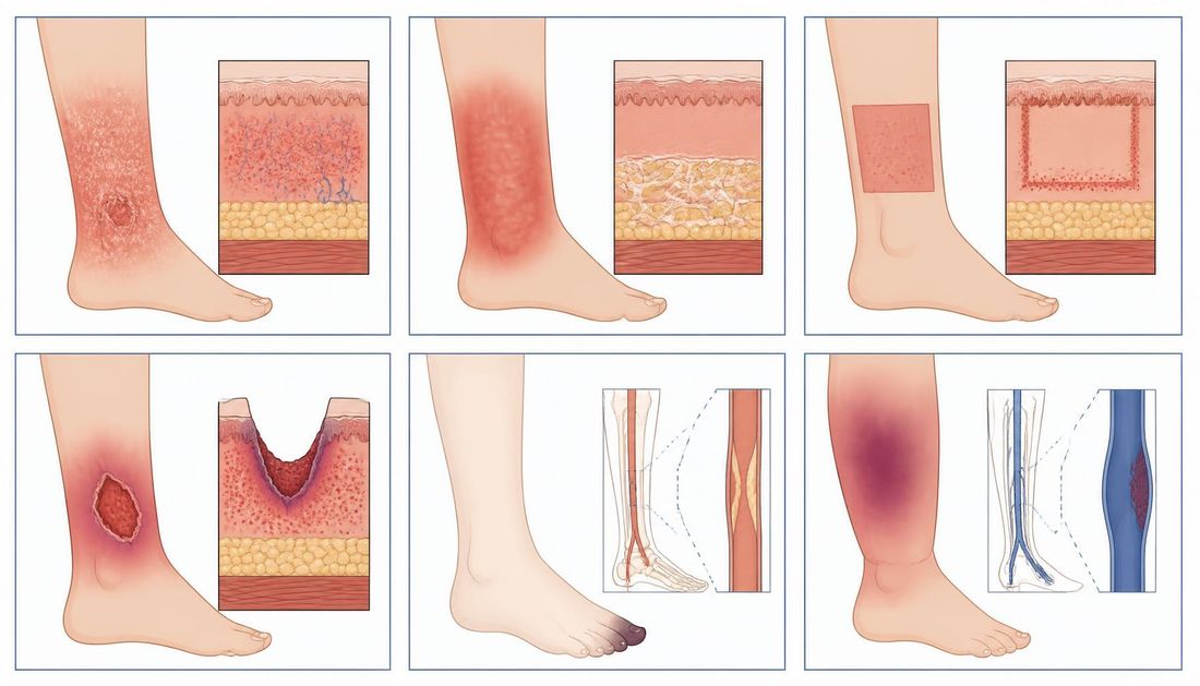
The six patterns shown provide a useful visual reminder: the distribution of the lesion, its border and its relationship to the shape of the dressing or to arterial perfusion often indicate the cause more effectively than skin colour alone.
For the diabetic foot, the joint 2023 guidelines from the International Working Group on the Diabetic Foot (IWGDF) and the Infectious Diseases Society of America (IDSA) strongly recommend diagnosing infection clinically, based on local or systemic signs of inflammation, and assessing its severity using a standardised classification. If the clinical examination is equivocal or difficult to interpret, inflammatory biomarkers may help as an adjunct, but they do not replace clinical assessment.
Why the same microorganism produces different presentations in two people
The same species and a similar microbial burden may have no consequences in one wound but cause spreading infection in another. The difference lies in the host and local conditions. Reduced arterial perfusion limits the delivery of immune cells, oxygen and medication; oedema and venous hypertension sustain inflammation and maceration; unrelieved pressure and shear continually cause further tissue damage.
Systemic factors act simultaneously. Poorly controlled diabetes alters neutrophil function and the microcirculation, neuropathy masks pain as an early sign, immunosuppressive and corticosteroid therapy attenuates the inflammatory response, and protein–energy malnutrition reduces tissue repair and immune reserve. Chronic kidney disease, active malignancy and advanced age further favour a clinically quiet presentation.
There are two practical implications. In patients with an attenuated response, the threshold for taking subtle changes seriously must be lower, because classic signs may remain absent until the process is already deep-seated. At the same time, when a patient's wound is stalled without signs of infection, the first question is not which antimicrobial product to add, but which of these factors has not been corrected.
Biofilm: an important concept without a simple diagnostic shortcut
A biofilm is an organised community of microorganisms held together by an extracellular matrix and characterised by altered cellular physiology. One of its most important properties is increased tolerance to antimicrobial agents and host defence mechanisms. Tolerance is not the same as genetically mediated antimicrobial resistance: cells within a biofilm can survive exposure to an antimicrobial agent without the permanent genetic change that defines conventional resistance.
However, biofilm in clinical practice is not a ‘slimy layer’ that can be reliably identified with the naked eye. The EWMA document cautions that much of our knowledge of biofilm comes from laboratory models that cannot fully reproduce the microenvironment of a chronic wound, and therefore warns against uncritically applying laboratory results to clinical wounds.
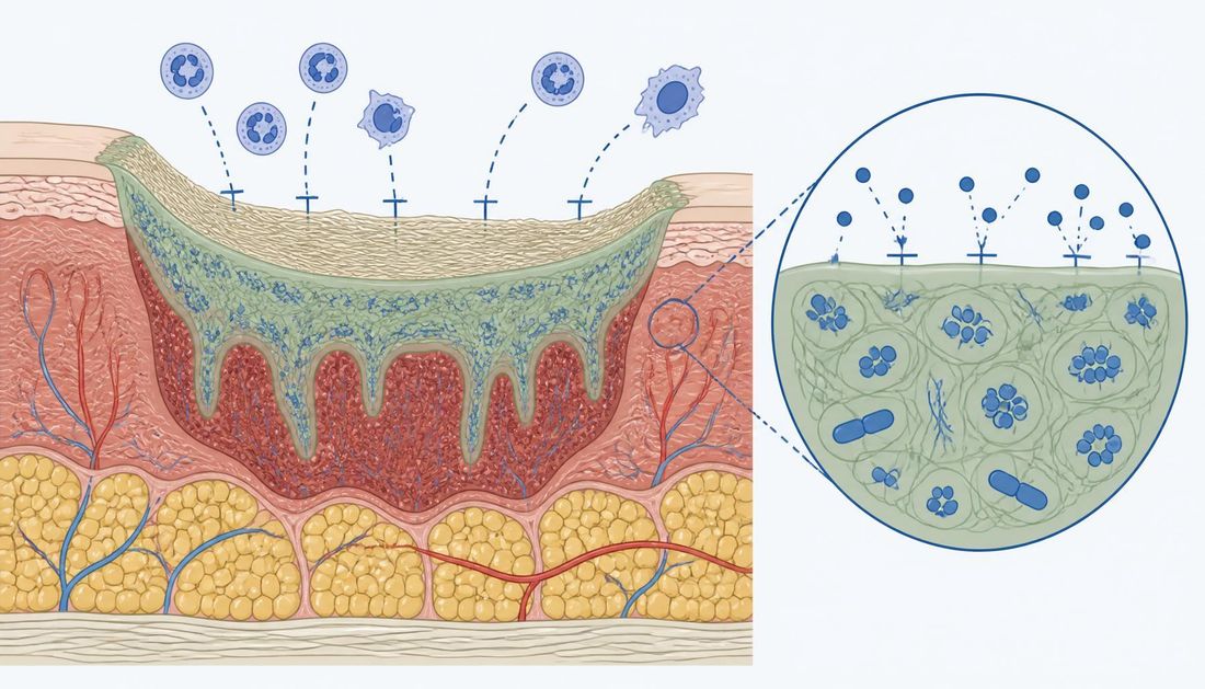
As shown in the cross-section, the matrix is a physical and physiological barrier: some of the antimicrobial agent is retained at its surface, while cells in the deeper layer reduce their metabolic activity and survive exposure without a permanent genetic change.
This has two practical implications. First, not every stalled wound should automatically be labelled as having ‘biofilm’ without reassessing perfusion, oedema, pressure, aetiology, nutrition and other causes of failure to heal. Second, if biofilm is suspected, mechanical reduction of devitalised tissue and microbial burden through cleansing and appropriate debridement is often a more important part of the strategy than endless rotation of antimicrobial dressings. IWII includes cleansing, debridement and biofilm-based wound care alongside antimicrobial stewardship principles in contemporary infection management.
A reduction in bacterial count is not the same as clinical cure of infection. EWMA emphasises that, because of the heterogeneous distribution of bacteria, quantitative monitoring of microorganisms by swab or even biopsy is not a universally reliable outcome measure for antimicrobial therapy. More clinically relevant outcomes are resolution of infection, wound improvement and, where appropriate, wound closure.
When and how to obtain a microbiological sample
A microbiological sample is useful when the result will change the treatment decision. There is no need to routinely swab every chronic wound simply because it is open. IWII lists spreading or systemic infection, failure of previous antimicrobial therapy and situations requiring surveillance of resistant microorganisms as appropriate indications.
Before sampling, surface debris should be removed and the wound appropriately cleansed to reduce the risk that the laboratory primarily identifies surface colonisers. The EWMA algorithm makes the presence of clinical infection the initial decision point, followed only then by microbiological sampling and selection of empirical therapy.
For suspected diabetic foot infection, IWGDF/IDSA prefers an aseptically collected tissue sample obtained by curettage or biopsy to a superficial swab. When osteomyelitis is suspected, a bone sample is preferred for microbiological diagnosis when sampling is justified and feasible.
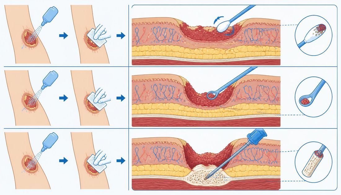
The illustration of three sampling methods explains why the results cannot be interpreted in the same way: a swab taken after cleansing reflects the surface, tissue curettage or biopsy reflects the wound bed, and a bone sample addresses the question of bone involvement.
When empirical therapy is needed for a serious infection, it should not be delayed solely to await microbiological results. Once results are available, the aim is to move from empirical therapy to the most precisely targeted treatment possible, according to the identified pathogen, susceptibility, infection severity, site of infection, patient safety and local epidemiological data.
Osteomyelitis: when to suspect it and how to confirm it
Bone involvement changes both the diagnostic approach and the duration of treatment, so it should be actively sought rather than discovered incidentally. Suspicion is increased by an ulcer that persists beyond the expected time despite appropriate care and offloading, an ulcer extending deeper than the subcutaneous tissue, exposed bone or tendon, a toe that is red and swollen throughout, and recurrent infection at the same site.
The probe-to-bone test, performed with a blunt sterile probe, is a simple clinical procedure that substantially increases the likelihood of osteomyelitis in a high-risk population, but is not proof on its own. Plain radiography is useful as a first step and may show bone destruction, a periosteal reaction or a sequestrum, but early findings are non-specific and a normal image does not exclude the process. When clinical suspicion remains high, magnetic resonance imaging has the greatest sensitivity for early bone and soft tissue changes.
Confirming the causative organism and planning treatment
For microbiological diagnosis, an aseptically obtained bone sample is valuable, rather than a swab of the overlying wound; surface findings often do not correspond to the causative organism in the bone. Bone sampling is useful when the result changes a decision — when the causative organism is unclear, previous therapy has failed, a resistant microorganism is suspected or prolonged antimicrobial treatment without resection is being planned.
Treatment is planned jointly with a surgeon: removal of infected bone shortens the required duration of antimicrobial therapy, whereas a conservative approach involves treatment planned over weeks, with clear criteria for success. The presence of osteomyelitis does not justify neglecting perfusion, offloading and local wound care; these remain prerequisites for healing regardless of the chosen strategy.
Antiseptics, topical antibiotics and systemic antibiotics are not the same
The term ‘antimicrobial agent’ encompasses different groups. Antibiotics are medicines that can be administered topically or systemically, whereas antiseptics are topical agents with broad antimicrobial activity. The distinction is not merely terminological, because indications, evidence, resistance risk, cytotoxicity and the intended therapeutic goal differ.
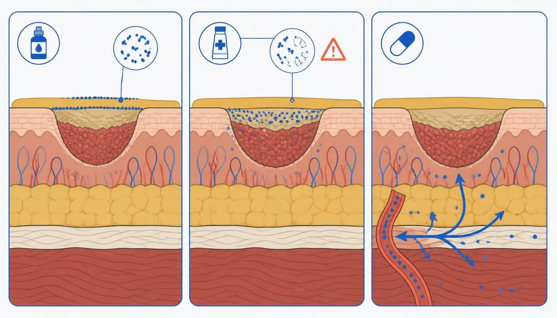
The illustration of different levels explains why these groups are not directly interchangeable: an agent acting on the surface does not reach the tissue where spreading infection is occurring, while a systemic medicine does not address the surface microbial burden and devitalised tissue that require mechanical removal.
Routine long-term use of topical antibiotics in chronic wounds is not justified. The EWMA document warns of overuse, allergic reactions and selection pressure for resistance. For diabetic foot infection, IWGDF/IDSA also conditionally recommends against adding topical antibiotics to systemic antibiotics for the treatment of soft tissue infection or osteomyelitis.
The role of antiseptics and the limits of a local approach
The situation is more nuanced for antiseptics. IWII gives them a role in locally infected wounds and selected high-risk situations, but selection should take account of the expected pathogens, antimicrobial spectrum, cytotoxicity, irritation, allergenic potential, treatment duration and local guidelines. Evidence that an antiseptic kills microorganisms in the laboratory is not, in itself, evidence that it accelerates healing in a clinical wound.
In spreading infection or systemic illness, a topical antimicrobial dressing cannot replace systemic therapy and source control. For the diabetic foot, IWGDF/IDSA specifically identifies the need for early surgical assessment in severe infection or moderate infection complicated by gangrene, necrotising infection, a deep abscess, compartment syndrome or severe ischaemia.
Systemic therapy: route of administration, duration and de-escalation
For mild soft tissue infection in a patient with adequate absorption who can take medication by mouth, the oral route is usually sufficient. The parenteral route is preferred in severe infection, a marked systemic response, unreliable absorption, inability to take oral medication or when the expected pathogen requires a concentration that an oral preparation cannot provide. Once the patient's condition has stabilised and the infection is clearly resolving, switching to oral therapy reduces the need for hospital-based interventions without loss of efficacy.
Duration is determined by the clinical response and extent of the process, not by whether the wound has closed. For infection confined to soft tissue, treatment is planned in days rather than weeks and ends when the signs of infection have resolved. When bone is involved, treatment is longer and depends on whether the infected bone has been surgically removed or is being treated with antimicrobials. Continuing therapy solely because the wound has not yet epithelialised constitutes exposure without expected benefit.
Empirical selection and de-escalation
Empirical selection is based on the expected pathogens, infection severity, local epidemiology, previous antibiotic exposure, allergies, renal and hepatic function, and interactions with existing medication. For a mild superficial infection without risk factors, coverage of aerobic Gram-positive pathogens is usually sufficient; a broader initial spectrum is reserved for severe infection, previous antimicrobial therapy, known colonisation with a resistant microorganism or extensive tissue damage.
De-escalation is part of the plan, not an afterthought. Once microbiological findings and susceptibility results are known, therapy is narrowed to the most appropriate narrow spectrum; when the findings do not explain the clinical presentation, the representativeness of the sample and the accuracy of the diagnosis are reassessed. A pre-agreed review date prevents treatment from continuing through inertia.
Antimicrobial stewardship: starting treatment appropriately and knowing when to stop
Antimicrobial stewardship is not a strategy of ‘as few antibiotics as possible at any cost’. The aim is for patients with infection to receive timely, effective treatment, while those without infection are not unnecessarily exposed to antibiotics. EWMA identifies the key principles as avoiding treatment when it is not needed, selecting the appropriate class and route of administration, using the narrowest appropriate spectrum and treating for the shortest duration required for clinical cure.

As the circular diagram shows, the four principles are not separate: the decision to start therapy determines the spectrum, the spectrum determines the duration, and reassessment brings the cycle back to its starting point. The symbols around the circle are reminders that none of these steps replaces drainage, debridement, revascularisation or offloading.
This message is even more important today. In July 2026, the World Health Organization reported that bacterial antimicrobial resistance was associated with more than 4.7 million deaths worldwide in 2021, and that approximately one in six laboratory-confirmed bacterial infections monitored through global surveillance in 2023 was resistant to antibiotics. Inappropriate and excessive antibiotic use is listed among the main drivers of resistance.
For wounds, this means that antimicrobial therapy must be linked to management of the underlying cause. An antibiotic cannot replace abscess drainage, removal of necrotic tissue, revascularisation of an ischaemic limb, offloading of the diabetic foot or control of venous hypertension. IWGDF/IDSA explicitly warns that treating infection alone without appropriate local wound care, offloading, vascular assessment and metabolic control increases the risk of failure.
Reassessment when there is no response
Antimicrobial stewardship also includes re-evaluation. If an infection does not respond as expected, the question is not simply whether a stronger antibiotic is needed. It is necessary to reassess whether the diagnosis is correct and whether there is an inadequately drained collection, necrosis, osteomyelitis, a foreign body, ischaemia, a resistant pathogen or inadequate adherence to treatment. For diabetic foot infection, IWGDF/IDSA recommends reassessment if signs of infection have not resolved after appropriate therapy.
Razina dokazaHeterogeneous — expert review, international consensus and recommendations using GRADE (Grading of Recommendations Assessment, Development and Evaluation, a system for rating the certainty of evidence and strength of recommendations)EWMA / Journal of Wound Management 2022, IWII 2022, IWGDF/IDSA 2023The EWMA document is an expert review and practical update, and its authors explicitly state that it is not a formal clinical guideline. IWII 2022 is an international consensus, whereas IWGDF/IDSA 2023 uses GRADE and provides formally graded recommendations. The certainty of individual statements therefore varies, particularly for topical antimicrobial agents.
Before prescribing antimicrobial therapy
- Are there clinical signs of infection, rather than merely a positive microbiological result?
- Has a condition mimicking infection been excluded (venous and contact dermatitis, lipodermatosclerosis, inflammatory dermatosis, ischaemia, haematoma)?
- Have the severity and extent of the process been assessed: local, spreading, systemic, deep tissue or bone involvement?
- Is there a focus requiring source control — a collection, necrosis, a foreign body or ischaemia?
- If sampling is indicated, was the sample obtained after cleansing and from representative tissue?
- Have the treatment goal, route of administration, expected duration and review date been defined?
- Is de-escalation planned once microbiological results are available?
- Is the wound aetiology being addressed at the same time — compression therapy, offloading, perfusion, oedema and nutrition?
Clinical decision algorithm
Postupak
Chronic wound infection — from suspicion to targeted therapy
- 1. korak: If the wound has no clinical signs of infection, do not prescribe an antibiotic simply because it is chronic or microbiologically colonised; if signs are present, proceed to assess severity.
- 2. korak: If only one non-specific sign is present, look for additional clinical indicators and alternative diagnoses; if there is a combination of compatible signs, infection becomes more likely.
- 3. korak: If there is systemic deterioration, rapid spread of the process, necrosis, a deep abscess or severe ischaemia, arrange urgent assessment and source control; if not, determine whether the infection is local or spreading.
- 4. korak: If classic signs are attenuated by neuropathy, ischaemia, immunocompromise or older age, look for subtle signs such as an unexpected stall in healing, friable granulation tissue, increased exudate and new pain.
- 5. korak: If the clinical findings are compatible with dermatitis, venous inflammation, ischaemia, inflammatory dermatosis or another non-infectious condition, resolve the differential diagnosis before escalating antimicrobial therapy.
- 6. korak: If a microbiological result will not change treatment, do not routinely obtain a sample; if it will guide therapy or the infection is not responding to treatment, obtain an appropriate sample after wound cleansing.
- 7. korak: If the infection is serious, start the necessary empirical therapy according to severity and local guidelines without dangerous delay; if the infection is mild and superficial, tailor the strategy to the clinical situation.
- 8. korak: If there is an abscess, necrosis, a foreign body, a deep infected space or another surgically remediable barrier, undertake source control; if not, continue conservative treatment with close monitoring.
- 9. korak: If local perfusion is inadequate, incorporate vascular assessment and treatment; if perfusion is adequate, continue treatment of the underlying cause and local wound care.
- 10. korak: Once microbiological results are available, narrow therapy according to the relevant pathogen and susceptibility where possible; do not treat colonisers without a clinical indication.
- 11. korak: If clinical signs are decreasing, continue only for as long as necessary according to the type and depth of infection; if the expected response is absent, do not automatically prolong the same treatment without reassessment.
- 12. korak: After the infection has resolved, discontinue antimicrobial therapy when it is no longer indicated and refocus on treatment of the underlying cause, prevention of recurrence and the wound-healing plan.
The original EWMA algorithm follows the same fundamental logic: first ask whether clinical infection is present, and only then decide on culture, empirical therapy, interpretation of results and the definitive antimicrobial regimen. This is more important than any list of individual products.
When it is urgent: immediately, within 24 hours and within 72 hours
Immediate assessment is required for patients with suspected sepsis, shock, rapidly progressive necrotising infection, extensive gangrene, sudden worsening of ischaemia, neurological deterioration or another life- or limb-threatening condition. Spreading infection may involve deep tissue, fascia and muscle, while systemic infection may lead to organ dysfunction.
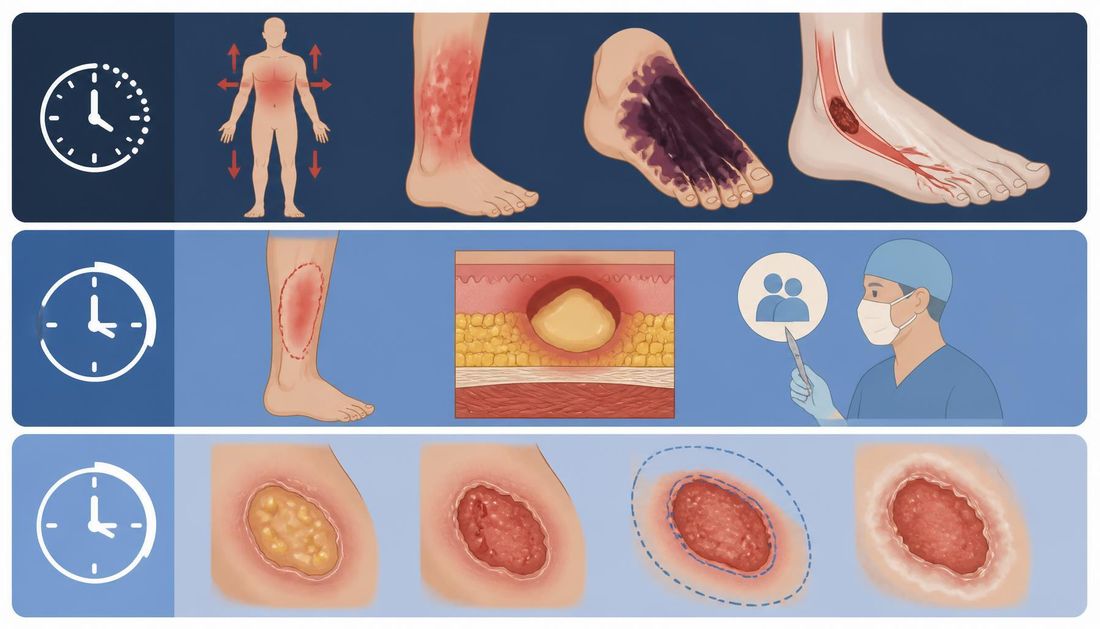
The bands in the diagram distinguish time frames, not just severity. The upper band describes conditions in which delay affects survival or limb preservation. The middle band covers conditions requiring specialist and surgical assessment within one day. The lower band describes a deteriorating trend that does not yet meet emergency criteria but requires earlier reassessment.
Serious infection or infection with progressive cellulitis, suspected deep abscess, necrosis, gangrene or significant arterial ischaemia requires specialist assessment within 24 hours. For diabetic foot infection, IWGDF/IDSA recommends urgent surgical consultation for severe infection or moderate infection complicated by extensive gangrene, necrotising infection, a deep abscess, compartment syndrome or severe ischaemia, and consideration of early surgery within 24 to 48 hours for selected moderate and severe infections.
New local pain, increased exudate, unexpected wound enlargement, newly friable granulation tissue, progressively worsening malodour, new maceration or another deteriorating trend that does not yet meet emergency criteria should be reassessed within approximately 72 hours. This interval is a practical triage framework for this article; it is not a universal formal time limit for every chronic wound.
Common errors in practice
Prescribing antibiotics because of a positive swab. This results in treatment of colonisation, unnecessary selection pressure and potential adverse effects. The correct approach is to link antibiotic use to clinical infection and use microbiological findings to guide therapy.
Labelling every stalled wound as having ‘biofilm’. This leads to neglect of ischaemia, oedema, mechanical loading or an incorrectly identified aetiology. The correct approach is first to reassess the patient and wound as a whole.
Rotating antiseptics without a goal or review date. This may result in unnecessary exposure, irritation or delayed treatment of the underlying cause. The correct approach is to define in advance why a topical antimicrobial approach is being used and when its effect will be evaluated.
Errors in treatment decisions
Neglecting source control. An antibiotic does not drain an abscess or remove devitalised tissue. When a surgically remediable focus is present, antimicrobial therapy must be part of a broader strategy, not a substitute for it.
Errors in monitoring and measuring success
Overly broad empirical therapy without de-escalation. Once relevant microbiological results are available, treatment should, where possible, be tailored to the actual causative organism and local guidelines. This is fundamental to antimicrobial stewardship.
Measuring success solely by a ‘cleaner swab’. The clinical goal is resolution of infection and improvement in the patient and wound, not sterilisation of a chronic wound.
Frequently asked questions
Česta pitanja
Does every chronic wound require microbiological testing?
No. Sampling is most useful when clinical infection is present and the result may influence therapy, when treatment has failed or in other clearly defined situations. A colonised wound without clinical infection does not require a routine swab.
Does Pseudomonas aeruginosa in a swab mean a systemic antibiotic is needed?
Not automatically. The microorganism must be interpreted alongside the clinical findings and sample quality. IWGDF/IDSA even recommends against routinely providing empirical coverage for P. aeruginosa in every diabetic foot infection in temperate climates.
Is malodour sufficient to diagnose infection?
No. Increased malodour may be one sign of local infection, but diagnosis is based on a combination of clinical signs and the overall condition of the wound and patient.
Can an antiseptic replace an antibiotic?
Not in spreading or systemic bacterial infection that requires systemic treatment. A topical antiseptic has a different role, primarily in the local management of selected wounds.
Should antibiotics be given prophylactically because a wound has not healed for a long time?
Not solely because the wound is chronic. For a clinically uninfected diabetic ulcer, IWGDF/IDSA explicitly recommends against using antibiotics to prevent infection or accelerate healing.
What is most important if an infection is not responding to treatment?
Recheck the diagnosis and source control: is there a deep abscess, necrosis, osteomyelitis, a foreign body, ischaemia, an inadequate sample or a pathogen not covered by treatment? Escalating antibiotics alone is not a sufficient strategy.
Izvori
References
Popis izvora korištenih pri pisanju ovog članka.
1. izvor: Probst S, Apelqvist J, Bjarnsholt T, Lipsky BA, Ousey K, Peters EJG. Antimicrobials and Non-healing Wounds: An Update. Journal of Wound Management. 2022;23(3 Suppl 1):S1–S33
DOI: 10.35279/jowm2022.23.03.sup01
Otvori izvor (vanjska poveznica)2. izvor: International Wound Infection Institute. Wound Infection in Clinical Practice: Principles of Best Practice. International Consensus Update 2022
Otvori izvor (vanjska poveznica)3. izvor: Senneville É, Albalawi Z, van Asten SA, i sur. Guidelines on the diagnosis and treatment of foot infection in persons with diabetes: IWGDF/IDSA 2023
Otvori izvor (vanjska poveznica)4. izvor: World Health Organization. Antimicrobial resistance. Ažurirano 16. srpnja 2026
Otvori izvor (vanjska poveznica)5. izvor: World Health Organization. Global antibiotic resistance surveillance report 2025
Otvori izvor (vanjska poveznica)